-
|
| .
|

Junior Member
- Group
- Administrator
- Posts
- 2
- Status
- Offline
|
|
 Evaluation of three mummified animals ?a a cat, a hen in addition to a snake ?a from Historic Egypt employing advanced 3D X-ray imaging is explained in a very paper published in Scientific Reports. The method presents insights in to the disorders in which the animals ended up held, their sophisticated mummification course of action as well as their feasible causes of dying, without having resulting in injury on the specimens. Evaluation of three mummified animals ?a a cat, a hen in addition to a snake ?a from Historic Egypt employing advanced 3D X-ray imaging is explained in a very paper published in Scientific Reports. The method presents insights in to the disorders in which the animals ended up held, their sophisticated mummification course of action as well as their feasible causes of dying, without having resulting in injury on the specimens.
Richard Johnston and colleagues applied non-invasive X-ray microCT imaging to reveal the skull in the cat to be close to 50 % the scale with the exterior mummified wrappings. Its morphology suggests that the continues to be probable belong to an Egyptian domestic cat. Examination of photographs on the teeth and skeleton reveal which the cat was considerably less than five months aged and may have purposefully experienced its neck broken with the time of death or during the mummification system to help keep the head in an upright placement.
A few mummified animals from historical Egypt are already digitally unwrapped and dissected by scientists utilizing high-resolution 3D scans that give unprecedented element concerning the animals’ lives - and deaths - more than 2000 yrs ago. The a few animals - a snake, a fowl along with a cat - are in the collection held because of the Egypt Centre at Swansea College. Preceding investigations had recognized which animals they have been, but hardly any else was recognised about what lay in the mummies. Now, owing to X-ray micro CT scanning, which generates 3D visuals with a resolution 100 situations better than the usual medical CT scan, the animals’ continues to be is usually analyzed in incredible detail, proper right down to their smallest bones and teeth. Credit history: Swansea College
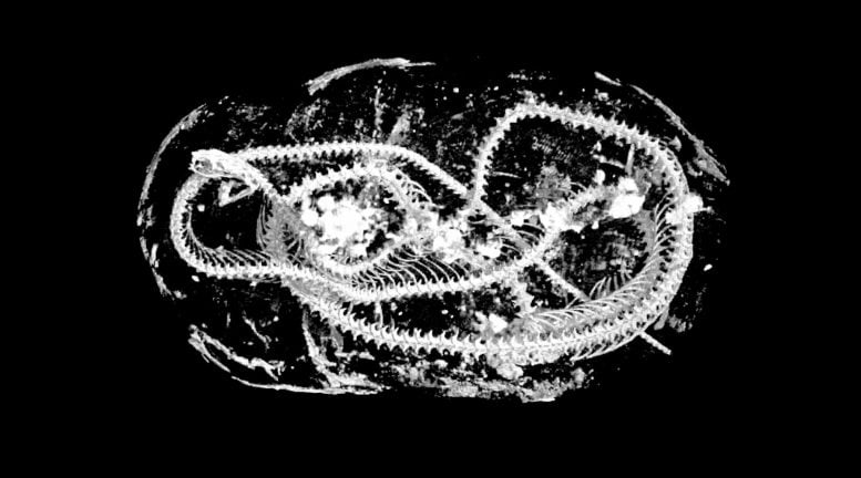
Measurements taken employing 3D scans on the mummified fowl of prey recommend which the continues to be most carefully resemble the Eurasian kestrel which the animal did not look to get died from accidents towards the neck. Imaging from the tightly coiled snake implies the stays belong to a juvenile cobra, which may have been killed by spinal fracture, dependable with tail capture and whipping strategies generally utilized to destroy snakes. The high-resolution imaging enabled the authors to determine structures found inside the mouth in the mummified snake as hardened resin. The exact placement with the opening from the glottis quite possibly presents evidence for intricate ritualistic behavior, much like the Opening with the Mouth method.
Digitally dissected reduced jaw (mandible) and enamel in the mummified kitten. Reveals fractures and unerupted mandibular initial molars (purple) indicating it had been a kitten in the time of loss of life. Scale: cranium whole duration = sixty eight.9 mm. The cat is one of three mummified animals from ancient Egypt which have been digitally unwrapped and dissected by researchers, making use of high-resolution 3D scans that give unparalleled detail concerning the animals’ life - and deaths - over 2000 several years back. Prior investigations experienced discovered which animals they were being, but little or no else was identified about what lay in the mummies. Now, because of X-ray micro CT scanning, which generates 3D visuals by using a resolution a hundred periods greater than the usual healthcare CT scan, the animals’ continues to be is often analysed in incredible detail, appropriate all the way down to their smallest bones and teeth. Credit: Swansea University
|
|
|
| .
|
0 replies since 4/9/2020, 10:09 3 views
.